
How does Cone Beam Computed Tomography work in 3D imaging?
In the realm of medical imaging, technological advancements continue to reshape the landscape, providing healthcare professionals with powerful tools for diagnosis and treatment planning. One such groundbreaking technology is Cone Beam Computed Tomography (CBCT), a remarkable imaging technique that offers three-dimensional insights into the human body. In this blog, we’ll delve into the intricacies of CBCT and unravel the mysteries behind its ability to create detailed 3D images.
Understanding the Basics:
Cone Beam Computed Tomography is a specialized form of computed tomography, commonly known as CT scanning. Unlike conventional CT scanners that use a fan-shaped beam, CBCT employs a cone-shaped X-ray beam. This unique configuration is particularly advantageous for capturing detailed images of a specific area, such as the head, neck, or extremities.
How CBCT Works:
-
Image Acquisition:
- The process begins with the patient positioned within the CBCT machine, which resembles a large donut or cube.
- The CBCT machine rotates around the patient, capturing a series of X-ray images from various angles.
-
Digital Reconstruction:
- The collected X-ray images are then processed digitally to create cross-sectional slices of the targeted area.
- These slices are high-resolution and provide detailed information about the internal structures.
-
Volumetric Rendering:
- The digital slices are compiled to reconstruct a three-dimensional volume of the imaged region.
- This volumetric rendering allows healthcare professionals to visualize intricate details in all dimensions.
Key Components of CBCT Technology:
-
X-ray Source:
- CBCT systems utilize a cone-shaped X-ray beam generated by a rotating X-ray source.
- This cone beam is designed to cover a specific anatomical area with precision.
-
Detector Panel:
- A detector panel opposite the X-ray source captures the transmitted X-rays after they pass through the patient’s body.
- The detector converts the X-rays into digital signals for further processing.
-
Data Processing Software:
- Sophisticated software algorithms process the digital data to reconstruct detailed 3D images.
- These images can be manipulated and viewed from different angles for comprehensive analysis.
Applications of CBCT in Healthcare:
-
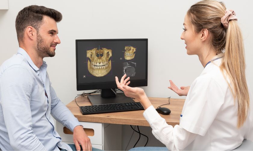
Dentistry:
- CBCT is widely used in dentistry for detailed imaging of the oral and maxillofacial regions.
- It aids in precise planning for dental implants, root canal treatments, and orthodontic procedures.
-
Orthopedics:
- CBCT helps orthopedic specialists visualize joints, bones, and soft tissues with exceptional clarity.
- It is valuable for pre-surgical planning and assessing musculoskeletal conditions.
-
Ear, Nose, and Throat (ENT):
- In ENT applications, CBCT assists in diagnosing conditions affecting the sinuses, nasal passages, and ear structures.
Conclusion:
Cone Beam Computed Tomography represents a remarkable leap forward in medical imaging technology, providing healthcare professionals with unparalleled insights into the human body. Its ability to generate detailed 3D images has revolutionized diagnostic and treatment planning across various medical fields. As CBCT continues to evolve, we can anticipate even more refined imaging capabilities, further enhancing its impact on healthcare outcomes.